Wednesday, October 31, 2012

Cardiac arrest

Cardiac arrest means the absence of contraction or inappropriate contraction of heart chambers immediately lead to the cessation of circulation of blood.


The cause of
A large number of causes that lead to cardiac arrest, primarily to the ventricular fibrillation, bradyarrhythmia, asystole, ischemic heart disease, left ventricular hypertrophy, mikarditisi, insufincijencija heart, conduction system disease, and others.

The clinical picture
Cardiac arrest is characterized by sudden loss of consciousness due to cessation of cerebral circulation.Cessation of respiration always follows the cessation of circulation and are usually recorded several deep respirations before the cessation of breathing. There is cyanosis (pomodrelost) A deepening. Pulse can not be felt beyond the pupil and after 1 minute the part, and after 2 minutes fully dilated. Time of termination of the circulation to vital functions are preserved at the level of cells known as clinical death. After 4 to 6 minutes caused irreversible brain damage and biological death.

Diagnosis
The patient was pulseless over large blood vessels, no heartbeat and no pressure, skin color is pale or cijanotična, respiratory movements are absent. The mechanism of cardiac arrest (ventricular fibrillation, asystole or bradyarrhythmia, elektromehaniča dissociation) can not be distinguished without ECG registration. All patients who survived cardiac arrest should be careful clinical examination, which, in addition to medical history and physical examination, including a 24-hour ECG test, the test load may exertion, ultrasound and coronary angiography.


Treatment

Should immediately start cardiopulmonary resuscitation. CPR is performed by blowing air exhaled by the resuscitator mouth to mouth or mouth to nose. Lips patients are cleared of foreign content, they are placed over tissue or something like that. Reanimator is with the patient, one hand underline under the door (and slightly below), neck lifts patient's head spontaneously falls back below the vertex towards the back. Use your thumb and index finger close nostrils second hand, palm of the same hand pressure head and pushes her head down as much as possible and back to the chin take the top position. A deep breath, put a mouth to mouth, and the patient blows air making sure that the coverage of your lips lips covered volume of patients, because it avoids the loss of air. Then reanimator frees patients mouth to allow air to uduvani out spontaneously. The first ten blowing out quickly and repeatedly and then proceed to blow straight to the rhythm of normal breathing (12 - 14 min). Checking blowing performance is evaluated emergence of synchronous movement of the chest.

External heart massage should be started as soon as you stop or severely weakened heart. It is performed by a steady, strong pressure in the connection area of ​​the upper two thirds and lower third of the sternum (breastbone). The suppression of the sternum to the spine from 3.5 to 5 cm is transferred to the heart. The person who helps raise the patient's legs Reanimator to move more blood to the heart and brain. About 2 cm above the end of the breastbone to place the root of the palm of one hand over the other hand its roots. The arms are extended at the elbows and shoulders above the sternum, so as the body weight is transferred to the resuscitator of his hand, it would be more efficient compression of the heart. Fingers placed his hands are not touching the chest. Hands are not separate areas of the sternum following compressions. Rhythm is 80-100 compressions per minute, and after every fifth compression is done once blow air at which point the compression is not performed. If resuscitation performed by two people, with the same rhythm of heart compression performed two uzastpona blowing every 15 compressions. Checking the performance is done after one minute of CPR. Signs of success CPR: palpated the pulse of the arteries in the neck or legs, dilated pupils are maximum (extended), improves your skin color, muscle tone occurs, the chest spontaneously and rhythmically driven.

Pulmonary artery stenosis

Pulmonary artery stenosis is an obstruction at the level of the right ventricular outflow tract and is relatively common congenital cardiac malformations. Narrowing be localized on the biota subvalvulnom level, at the level of the pulmonary artery and valve supravalvulno, and may represent a more localized combinations. Valve disease pulmonary stenosis is the most common form of isolated outflow tract obstruction of the right ventricle.


The cause of
Congenital heart defects occur in a complex interaction of genetic factors and environmental factors. It is rare to find the cause of congenital cardiac malformations such as, for example, maternal rubella or chronically excessive abuse of alcohol in the mother during embryogenesis. Various factors such as hypoxia, ionizing radiation, certain drugs have teratogenic effects. The risk of congenital heart defects in children of parents with congenital heart malformation is low, 2-5%, and would not need to be an obstacle in the planning of the offspring.

The clinical picture
Symptoms depend on the degree of narrowing of the pulmonary artery valve. Patients with mild stenosis usually have no symptoms and the disease usually does not show significant progress in the course of life, and reveal the findings of the noise survey. More severe stenosis prevents a corresponding increase in cardiac output during physical exertion and coming soon to the expression of fatigue, dyspnea (shortness of breath) and may lead to the occurrence of syncope (transient loss of consciousness). There may be opening up communication between the right atrium and left atrium at the level of the foramen ovale, which is the only significant part of the population is functionally closed, but not anatomically obliterisan. With the transition neoksigenisane blood from the right to the left prekomoru, and then the systemic circulation and leads to cyanosis (bruised). Like all cyanogen heart defects, and this slows down growth and increased body weight and height of the child. Greater sensitivity to the boys showed cyanosis.

Diagnosis
It is based on clinical presentation, physical examination (systolic ejection murmur), ECG, x-ray, ultrasound, cardiac catheterization and angiography.

Treatment
Surgical treatment associated with relatively small risk. Recently, more and more valve stenosis, pulmonary artery resolved without surgical intervention, cardiac catheterization laboratories, using a balloon catheter and performing balloon - catheterization.

Subarachnoid hemorrhage


Subarachnoid hemorrhage (SAH) is bleeding into the subarachnoid space, which usually occurs due to rupture of the aneurysm.


The cause of
Aneurysms are localized enlargement of the arteries. They may be congenital or may develop on the basis of congenital defect intima and media of the vessel, and then the ball - globular or spindle - fusiform. They are often localized to Wilisovom hexagonal and often there are more in the same person.Acquired aneurysms, whether on the basis of arteriosclerosis, diffuse are.

SAH may arise from arteriovenous malformations. Usually it comes tuft enhanced arterial and venous vessels with arterial input and output, venous vessels. By SAH can lead to the rupture of blood vessels wall defect without aneurysm. Blood will flow in suarahnoidni space at different speeds, depending on the size of the vessel that was ruptured but once the blood stream can damage the brain tissue.

The clinical picture
History is typical - Sudden head pain (like a punch), usually in the back of the head with the expansion of the neck, and sometimes down the spinal column to the lumbar regions and the frontal part of the head.Due to the rapid increase in speed occurs nausea, vomiting and disturbance of consciousness up to coma.

Because stimulus meninges meningeal signs appear, but they can sometimes be poorly expressed or absent. In case of damage to brain tissue can be found focal signs (hemiparesis half-body paralysis, seizures).

A very important finding is bradycardia (slow heart rate), which can be 40-50 beats per minute, and can serve as an indirect indicator of the size of the intracranial pressure.

The presence of blood in the subarachnoid space usually leads to vasospasm, which began to develop 3 - 4 days after bleeding, and lasts about three weeks. This serious complication of SAH leads to further, often diffuse brain damage becomes apparent reduction in the level of consciousness, mutism, or focal signs.

Prognosis depends on the severity of bleeding, the development and severity of vasospasm. Better prognosis in the absence of SAH aneurysm or AV malformation or with negative angiographic findings.Mortality is high, and recurrences are common, especially in the first six months.

Diagnosis
It is based on clinical findings, lumbar puncture, fundus findings, angiography, CT (scan).

Treatment
Conservative treatment consists of rest in the strictest (the patient may not even get up for their physiological needs). By all means necessary to prevent unrest (neuroleptics, benzodiazepines) and seizures (phenobarbital sodium, diazepam, parenterally), since they can cause new bleeding. Blood pressure control is also very important measure of compensation and 2-3 liters of fluid per day. Since arrhythmias are very common and neurogenic pulmonary edema can occur very early cardiopulmonary monitoring is necessary. Sometimes it is useful careful lumbar puncture and hipervolemic hemodilution using dextran. Hyperosmolar solutions can reduce brain edema and reduce inrakranijalni pressure.

It is very important issue when it comes to surgical intervention, and in which patients. The general agreement is that it can intervene before the development of vasospasm, ie. the first three days, in patients who are not in a coma. The others should wait for the withdrawal of vasospasm before the start artériographie studies and planning intervention, which is usually about three weeks.

Mitral stenosis


Mitral stenosis is a narrowing of the mitral defect caused the hole (the opening between the left atrium and ventricular) which lead to difficulty in emptying the left atrium into the left ventricle.


The cause of

Rheumatic endocarditis is almost exclusive cause of mitral stenosis, mitral stenosis is extremely rare congenital malformation is the result of the mitral valve.

The clinical picture

Patients with moderate to tolerate any effort. Only the presence of precipitating factors (fever, greater effort, anemia, atrial fibrillation, pregnancy) leads to shortness of breath - dyspnea and cough. In moderate forms and less effort leads to these symptoms manifest themselves in severe and at rest. At an advanced stage dominated by fatigue, which increases due to right heart decompensation, with the advent of liver enlargement and edema prifernih - island. The emergence of palpitations - a racing heart caused by frequent atrial arrhythmias and atrial fibrillation later. Cause arrhythmias and emboli in the systemic circulation (brain, limbs, abdominal organs). Less commonly, symptoms appear in the form of angina pectoris, pulmonary embolism or hoarseness. When examining the dominant means of peripheral cyanosis (blue fingers, ears, nose) with cheeks flushed cheeks giving a typical picture-"facies mitralis". Jugular veins are swollen due to right heart decompensation.


Complications

Possible complications of acute pulmonary edema, atrial fibrillation, systemic embolism, bacterial endocarditis, lung damage.

Diagnosis

It is based on history, physical examination, physical findings (highlighted first tone, audible otvartanje mitral valve, noise), x-ray of the heart and lungs (configured mitral heart), ECG, fonokardiografije, ultrasound, cardiac catheterization.

Treatment

Medical treatment consists of prevention of rheumatic fever and infective endocarditis. In pulmonary congestion given diuretics, the restriction of salt intake. In the case of atrial fibrillation should be given digitalisne preparations, verapamil or beta blockers. The appearance of embolism requires anticoagulation. Medical treatment can reduce stenosis and surgical treatment is applied - different ways of expanding narrowed mitral opening or installing artificial valve.

Tetralogy of Fallot


Tetralogy of Fallot is the most common congenital heart defect cijanozna and represents 10% of all congenital anomalies. Means the combination of: 1) the defect međukomorske compartments, 2) riding aorta, which is an opening above the defect to receive blood from both chambers, 3) pulmonary stenosis (obstruction of the outflow tract of the right ventricle), and 4) right ventricular hypertrophy.


The clinical picture

Systemic hypoxemia (low oxygen levels) is the cause of most of the symptoms and signs in children and adults. Children lag behind in growth. During physical activity appear dyspnea (disturbed, shortness of breath), and dizziness, and relief appearances at zauuimanju characteristic position - squatting. Although there are forms of tetralogy acijanotični (pale Fallot), which can be found in adults, the majority of patients with this defect is characterized by cyanosis (blue discoloration) with maljičastim fingers and polycythemia (increased number of cells in the blood). Cyanosis can be of varying intensity. Attacks worsening cyanosis occur due to increased tone of excitement in the infundibulum (fear, crying) or taking drugs that increase myocardial contractility. Anoksemične crisis (crisis without oxygen) with sudden weakness, loss of consciousness, seizures, and even death, attributed to spasm - the deterioration of infundibular stenosis. Not seen in adults and in children are a sign of serious defects and indications for prompt surgical correction. Natural history of conservatively treated patients is poor.Frequent kompikacije such as heart attack and brain abscess, and the risk of infective endocarditis is great. Heart failure and arrhythmias are the most common causes of death in most patients until 30 appearances year.

Diagnosis
It is based on clinical presentation, examination, ECG, X-ray, ultrasound and cardiac catheterization.


Treatment

Cijanotički paroxysmal attacks is urgently treated by placing the child in the position with the knees to the chest, giving oxygen and opiates. With prolonged anoxic attacks should be included infusion and acidosis corrected bicarbonate. Once the required general anesthesia or giving beta blockers to stop the attack.
Surgical treatment is indicated in all patients. The ideal is a total correction of defects in the first years of life, which consists in closing the defect međukomorske barriers and reducing obstructions in the outlet of the right ventricle. Most operated almost normal lives, but some occur arrhythmias and sudden death.

Thrombophlebitis migrans

Migrans thrombophlebitis (phlebitis saltans) is a rare form of recurrent superficial thrombophlebitis, which can affect large and small veins throughout the body, and is often associated with other diseases.


The cause of
It is believed that this is allergy - hiperergijskoj vascular response to a variety of factors, eg. to local bacterial infection, tumor antigens, and the like. The disease is quite common in patients with cancer of the pancreas, lung and prostate cancer, leukemia, gout, lymphoma and trombangitis obliterans - a.

The clinical picture
Parts are usually thrombosed superficial veins in the legs, abdominal wall, thighs and arms, mostly in younger and middle godnina. Thrombosed veins and inflammatory part of the indurisan (hardened), and as a hardening and palpated as a pen or a rope, which is painful, and the skin over the vein edematous (swollen) and erythematous (red). With disease relapse and involvement of one hour, then the other veins, can last for months or years. The veins of the internal organs (spleen, mesentery) may also be affected, but it does not occur embolism.

Diagnosis
It is based on history, clinical, dermatological examination.

Treatment
Treatment is carried out Tylenol, the eventual systemic corticosteroids. Locally applied heparin and heparinoidni preparations. It is necessary to treat the local infection with antibiotics and stop smoking.

Raynaud's Syndrome


Raynaud's syndrome, intermittent episodic occurrence mutual pale and blue fingers, legs less often, because of occlusion (closing) digital arteries (arteries in the fingers) precipitated by cold or emotional stress, undergoing warming.


The cause of
It can be divided into primary or idiopathic, or secondary. Diseases related secondary Raynaud's syndrome are collagenosis (systemic sclerosis, systemic lupus erythematosus, rheumatoid arthritis, Sjogren's anchor, diabetes mellitus), and other vascular disease, obstructive disease of the arteries (arteriosclerosis, thrombosis, Morbus Buerger), poisoning (ergotaminski alkaloids, vinyl chloride, cytotoxic drugs, heavy metals), neurological and neoplastic processes, and heating professional or injury (frostbite injuries, vibration syndrome).

The clinical picture
Raynaud's syndrome affects between 3 and 5 million people worldwide, mostly women. It is characterized by local paleness, cyanosis, pain, a burning sensation or loss of sensation to the skin, swelling, and in extreme cases can result in unpleasant ulcers (ulcers) in the skin.

Diagnosis
It is based on history, clinical examination and confirmed by capillary microscopy nail beds (see the loss, and tortuosity of capillaries and venules extension).

Treatment

General measures include advice to avoid the cold, tobacco, excitement, physical exertion.Symptomatic treatment includes the use of vasodilatory drugs. Surgical treatment can lead to transient blood flow to the fingers. Infected ulcers should be treated with antibiotics.

Restrictive cardiomyopathy


Restrictive cardiomyopathy is characterized by impaired diastolic relaxation chamber, due to rigid walls of the heart chambers, thus, prevented them properly filling with blood.


The cause of
A large number of processes that cause infiltration and myocardial fibrosis can cause this disease. There are two forms of the disease: idiopathic, which is rare, and the secondary, whose causes are numerous: amyloidosis, sarcoidosis, endomyocardial fibrosis, Loefflerov hipereozinofilni endocarditis, carcinoid, and endocardial fibroelastoza hemochromatosis.

The clinical picture
Dominated by symptoms of heart failure, and often, right. It is located nabreklost neck veins, peripheral edema (swelling), hepatomegaly (enlarged liver), and sometimes ascites (presence of fluid in the abdomen). Often occurs dyspnea (shortness of breath), fatigue and edema. Heart sounds are quiet, there is tachycardia (rapid heartbeat) and frequent arrhythmia.

The disease is progredijentnog flow for several years, ending in death, mainly due to refractory heart failure.

Diagnosis
It is based on history, clinical findings, objective examination (tricuspid regurgitation murmur), ECG, x-ray, ultrasound and cardiac catheterization.

Treatment
Treatment is aimed at improving heart failure. Be conducted, with sparing patients the larger effort, the symptomatic treatment of heart failure with diuretics, digoxin, ACE inhibitors, antiarrhythmics. When the existence of the risk of systemic embolism should be given oral anticoagulant. Medical therapy is often ineffective, and sometimes need surgery consisting of excision of fibrotic endocardium and valve replacement.

Tricuspid stenosis


Tricuspid stenosis (stenosis of the right vein opening) in which a defect, tricuspid stenosis due to the opening, there is an obstruction in the movement of blood from the right atrium to the right ventricle.


The cause of
As acquired defects, most frequently caused by rheumatic fever, when associated with mitral stenosis.Less commonly tricuspid stenosis occurs within fibroelastoze endocardium, endomyocardial fibrosis, systemic lupus erythematosus or carcinoid syndrome.

Tricuspid stenosis as a birth defect caused by congenital abnormalities of the tricuspid valve in the heart of the complex malformations.

The clinical picture
The clinical picture depends on the support of the dominant defects (mitral, aortic). If expressed tricuspidal stenosis, dominated fatigue, dyspnea (shortness of breath) and pain in the liver area (venous stasis). Manifested clinical signs are: swollen neck veins, hepatomegaly (enlarged liver), ascites (fluid in the abdomen), peripheral edema (islands).

Diagnosis
It is based on history, clinical presentation, physical examination, ECG, radiology, ultrasound and cardiac catheterization.

Treatment
Diuretics to reduce systemic venous congestion is allowed in isolated and moderately severe forms of the disease. Surgical treatment is considered in the presence of concurrent anomalies and indications for such treatment arises in the case of reduction of tricuspid opening under 2 cm ², or the existence of a pressure gradient of 5 mmHg or more. Surgical treatment involves tricuspid valvuloplasty or valve replacement, and recently he performed balloon dilatation of the tricuspid orifice.

Thrombophlebitis


Means the occurrence of thrombophlebitis thrombus and inflammatory reaction of the walls of the superficial veins.


The cause of

Thrombophlebitis is common in the symptomatology of varices of the lower extremities, especially in females. The pathogenesis of thrombosis, the endothelium bit vein walls, slowing blood flow and blood hypercoagulability, all resulting in, for example. varices, venous trauma, prolonged lying in bed, using oral contraceptives, septic conditions and malignant infiltration of the walls of the veins.

The clinical picture

Inflammation (inflammatory) changes commonly observed in varicose veins on the thighs and lower legs. Thrombosed veins and inflammatory part of the indurisan (hardened), and as a hardening and palpated as a pen or a rope, which is painful, and the skin over the vein edematous (swollen) and erythematous (red). Body temperature is elevated. After a few days thrombophlebitis regress, and lags mass of connective tissue in the affected segment of vein (phlebosclerosis). Recurrences are common.
Diagnosis

It is based on history, clinical, dermatological examination.
Treatment

Apply Tylenol, salicylates, and platelet aggregation inhibitors, and antibiotics are indicated only for septic thrombophlebitis. Locally applied heparin and heparinoidni preparations. Diseased leg bends tourniquet. Be sure to repair all the factors that favor the development of thrombophlebitis.

Ventricular arrhythmias


Ventricular (ventricular) arrhythmias usually occur due to ischemia (myocardial infarction, angina pectoris, chronic coronary insufficiency, myocardial aneurysm, angina vazosppastic), the cardiomiopaty, myocarditis, long QT - interval, mitral valve prolapse, physical exercise, right ventricular dysplasia artimogene and others., and can occur without clinically apparent disease.


Ventricular premature beats (ventricular premature beats, VES) on ECG - to be recognized by a wide QRS ompleksu, which is not preceded by P wave, and the result is abnormal and start the flow, and delayed ventricular depolarization. Origin and appearance may be different (multifokalneVES), compared to the previous normal PQRST may occur early during repolarization, the previous talasuT (R on T phenomenon). VES can occur in late diastole and be cause fusion complex or interpolated between two normally conducted sinus impulses. When you arise after each sinus impulses, it is a bigeminy, and after two pulses of trigeminiji. Two consecutive heather are a couple, three or more ventricular tachycardia.

Many patients do not feel the appearance of VES, while others were of the mind skipping the heart work harder or heart attacks. If there are no symptoms, no other individual VES clinical myocardial injury does not require medical treatment, and the injured myocardium, especially in the damaged left ventricle VES, VES in pairs, can be the sign of multifocal malignant ventricular arrhythmias and require treatment. In addition to treating the underlying disease, apply for beta blockers and antiarrhythmics I and III groups.
Ventricular tachycardia (ventricular tachycardia, VT) is defined as a series of three or more premature ventricular beats at a frequency between 100 and 250/min. It occurs due to the existence of the level of ectopic center chamber with fast-frequency pulses of creation. Presenting ventricular complexes that are bizarre with prolonged QRS and diskordinatnom final oscillation. As occurs in patients with significant myocardial damage, it can cause or worsen heart failure or cardiogenic shock. The most commonly manifested as palpitations (heart palpitations), weakness, dizziness, and dyspnea (shortness of breath).


Treatment of symptomatic forms is not required. In patients with acute myocardial infarction intraveska lidocaine and mexiletina is very effective. Treatment involves correction of attack precipitating factors, and medication from a drug of first choice is intravenous lidocaine, an alternative to beta-blockers, antiarrhythmics and groups. If the drugs do not have a quick effect elektrokatdioverzija the treatment of choice.
Chambers flutter (flutter ventriculorum) is due to the level of focus ektopičkog myocardium whose function is 250-300/min. It is characterized by QRS complexes is extended in the form of regular sine waves without the possibility of recognition of T wave. Chambers flutter leads to pronounced hemodynamic changes that are reflected in declining cardiac output. Flutter often goes into fibrillation chamber, because he needed urgent treatment, which consists of priomene elektrocardioversion.
Flickering of Commerce (fibrillatio ventriculorum) is chaotic activation myocardium resulting from the existence of ectopic focus in the heart muscle chambers with resultant circular motion at the level of Purkinje fibers. Fibrillation is the cessation of heart. They are registered in multiple, irregular, and different apmlitude duration, sinusoidal ripples.


Treatment is urgent, normal heart activity can be established defibrillation chamber (DC shock), the following procedure was cardiac massage and artificial respiration, followed by intravenous lidocaine and epinephrine, or both drugs together

Ventricular septal defect


Ventricular septal defect (VSD) is the most common congenital heart malformations and can exist as isolated vicijum, or associated with other defects, as part of complex congenital heart defects. Usually there is an opening, located basal, level membranous part of the septum.


The cause of
Congenital heart defects occur in a complex interaction of genetic factors and environmental factors. It is rare to find the cause of congenital cardiac malformations such as, for example, maternal rubella or chronically excessive abuse of alcohol in the mother during embryogenesis. Various factors such as hypoxia, ionizing radiation, certain drugs have teratogenic effects. The risk of congenital heart defects in children of parents with congenital heart malformation is low, 2-5%, and should not be an obstacle in the planning of the offspring.

The clinical picture
The clinical picture depends on the size of the ventricular septal defect. Small defect is usually not accompanied by symptoms. Significant septal defect with left - right shunt causes a moderate degree of poor tolerance. Recirculation of blood in the pulmonary circulation reduced cardiac reserve, and the effort quickly and leads to weakness and fatigue. Large VSD leads to slowing growth and physical development of the child, the appearance of frequent respiratory infections, and can lead to cardiac failure due to high loads of left ventricular volume. After the second decade of life, there is a manifestation of pulmonary vascular obstructive disease with pulmonary hypertension and reduced left - right shunt. Bidirekcionog occurrence and, later, the right-to-left shunt causes cyanosis (bruised, blue discoloration) and causes a polycythemia (increased number of blood cells). Vascular obstructive pulmonary disease restricts blood flow increase in the effort and adequate increase in cardiac output as needed to load quickly and leads to fatigue and weakness, and may lead to the occurrence of syncope (transient loss of consciousness). Hypoxemia and inadequate increase in coronary flow during the effort, considering the needs of right ventricular hypertrophy, can trigger myocardial ischemia in the effort, and the occurrence of anginal chest pain. Pulmonary vascular obstructive disease and pulmonary hypertension can cause development of hemoptysis (coughing up blood).

Diagnosis
It is based on review (noise), ECG, x-ray, ultrasound and cardiac catheterization.

Treatment
VSD belongs to a group of congenital heart disease with a relatively high risk of bacterial endocarditis.Antibiotic prophylaxis of bacterial endocarditis is necessary in the case of interventions that could lead to bakteriemije (dental procedures, bronchoscopy, placement of urethral catheters, abortion, etc.).

Surgical treatment of VSD - also indicated in situations where a significant left to right shunt. In situations of existence odmaklih pulmonary vascular obstructive changes with markedly increased resistance and the emergence of reversion shunt surgical intervention is contraindicated septal defect closure. Alternative to surgical closure in some cases could be placed centrally Transcatheter closure of defects by using a special shutter as a double umbrella (clamshell). The optimal time for surgery has disadvantages preschool children, but the results are good, and operational risk is low even in patients older than 40 years if no pulmonary hypertension.

Vertebrobasilar insufficiency


Syndrome, disorders of blood circulation in vertebrobasilary - basilar system is defined almost half a century. Vertebral - basilar artery system with its branches supplying blood brain stem, cerebellum, occipital lobes and the temporal part of the hemisphere.


The cause of
The main causes of occlusive changes, most arteriosklerotske with thromboembolic changes (formation of aggregates of fibrin and platelets to complicated plaques, and their firing embolism of small blood vessels). In addition, you could be hypotension (low blood pressure), poor collateral circulation of the brain, neck rotation with compression of vertebral arteries in cervical spondylosis etc..

The clinical picture
Clinical syndrome vertebo - basilar (VB) failure are:

• Dizziness (vertigo) is the most common symptom of VB insufficiency. The patient has the impression that the cases revolve around him (objective vertigo) or sinking, falls (subjective vertigo). May be accompanied by nausea, vomiting, weakness.
• transient visual disturbances - flash, colors, parts outburst visual field hemianopia (half of visual field incidents), complete loss of vision. These disorders are due to ischemia in the posterior cerebral artery area
• ataxia (gait groggy)
• syncope (transient loss of consciousness), due to damage sinkopalnog reflex in the medulla oblongata
• "drop" Atacama - a sudden loss of strength in my legs and drop, without loss of consciousness due to ischemia pyramidal tract in the pons area
• diplopia (double vision)
• dysarthria (difficulty in pronunciation)
• transient cranial nerve damage
• transient deafness
• occipital headache
• transient global amnesia
• hemiparesis (paralysis of half the body)
• hemianestezija (loss of sense of touch in the middle of the body)
Sometimes one can see alternate side involvement, for example. in one attack - right-sided hemiparesis, in the second - dextral (seesaw phenomenon).

Diagnosis
It is based on history, clinical, neurological examination, fundus examination, laboratory tests (blood lipids, blood glucose, hemorrheological test), ultrasound, scintigraphy, CT scan (scanner) or NMR (nuclear magnetic resonance).

Treatment
Treatment of acute focal damage to one of the UK where failure is mainly aimed at the prevention of new attacks and ischemic stroke incidence complete. In the first place, we must fight against hypertension. The fight against smoking and alcohol may also contribute to the reduced number of patients. A healthy diet is an important factor in preventing vascular disease. It is primarily a food rich in fiber and vitamins (fruits, vegetables, herbaceous), food that relieves slow carbs and low fat foods of animal origin. Moderate physical (walking, working in the garden, etc.) and mental work are very useful.From medikamentaa be used in the prevention of vasoactive drugs, antiaggregational therapy (aspirin), calcium channel blockers. And where possible surgical treatment.

Wolf - Parkinson - White Syndrome


In the normal heart Atrial impulses can be conducted to the ventricles via the AV node only. Excitation (activation, which is earlier than normal) when there is an impulse from the atria bypasses the normal atrioventricular retention and rapidly cools the chamber. In preekscitacionim syndromes are akscesorni consignment roads connecting atrium and infranodalna fiber conduction system in which no holds pulses as in the AV node or connecting atrium and ventricle. Therefore Atrial conduct impulses much faster akscesornim roads and activation of Commerce begins earlier than the momentum going through the AV node to conduct commerce.

In patients with WPW syndrome are frequent attacks of paroxysmal supraventricular tachycardia (rapid heartbeat), which are mainly caused by rapid circular motion pulse frequency, which can first go through the AV node and then through akscesorni time or vice versa.

The cause of
WPW syndrome is a congenital anomaly. Familial occurrence has been described, but not a rule. Most patients, 80-90%, with WPW ECG image or WPW syndrome have a healthy heart. Once the WPW associated with congenital disorders such as Ebstein anomaly, mitral valve prolapse, or hypertrophic cardiomyopathy.


The clinical picture
Patients in the absence of an attack of paroxysmal tachycardia are asymptomatic. The symptoms of these attacks come from arrhythmia. The attacks are usually short-lived usually stop spontaneously, but can be interrupted and vagal stimulation. Some patients may have frequent and prolonged attacks that limit work capacity and reduced quality of life. A smaller percentage of patients may be rare, but expressed episodes quick re-entry tachycardia or atrial fibrillation leading to syncope (loss of consciousness). Rarely, the first and only event in a previously asymptomatic patients with WPW ECG image may be sudden cardiac death due to an attack of atrial fibrillation, which degenerate into ventricular fibrillation.

Diagnosis
WPW syndrome electrocardiogram (ECG) is characterized by a short PR interval, QRS complex, extending from the presence of delta waves in the ascending part of the R wave. In patients with atrial fibrillation can find rare irregular normal ventricular complexes and more frequent ventricular complexes with delta waves.

Treatment
When the attack of paroxysmal tachycardia should first try to stop the attack to stimulate vagal maneuvers. If the attack does not stop, give it some class IC antiarrhythmic drugs (ajmaline, flecainide or propafenone) or class IA (procainamide, quinidine or disopyramide). If the tachycardia stops elektrokardioverzija the method of choice. In patients with frequent attacks electrophysiological studies are needed that can localize Aberrant pathways and surgically severed.

Septic endocarditis


Definition:

Septic endocarditis entities include acute, subacute bacterial endocarditis, previously called endocarditis lenta, as well as other non-bacterial endocarditis caused by a virus or fungus. The disease is a significant cause of morbidity and mortality in children, despite progress in treatment and prophylaxis.


Etiopathogenesis:

For the development of infective endocarditis in 50% of cases responsible Streptococcus viridans. In 30% of cases, with increasing frequency in recent decades, is responsible Staphylococcus pyogenes. The participation of other organisms is less common and 10% of blood cultures were negative.Staphylococcal endocarditis is less common in patients who have primary heart disease. Streptococcus (viridans) endocarditis occurs after dental surgery, and enterokokni (group D) after manipulation of the genitourinary tract and GIT. Pseudomonas aeruginosa occurs after IV drug use.

Endocarditis is the most common complication of congenital or rheumatic heart disease, but can also occur in those children who have no previous heart disease. The disease is extremely rare in the infant age. Vegetation is usually formed at the site of intimal or endocardial lesions are the result of turbulent blood flow high speed and thus, the greatest risk for the development of septic endocarditis having children with: VSD, valvular stenosis, especially the aorta, tetralogy of Fallot, patent ductus arteriosus, transposition of the great right vascular and surgical systemic-pulmonary shunts. In adults, this can add up bikuspidalna aortic valve, mitral valve prolapse with mitral regurgitation, the artificial valve.Surgical correction of heart disease reduces but does not eliminate the risk of endocarditis, except repair 6 months after ASD closure and DAP. Increased risk of developing this disease are drug users, and those who have a long-term presence of catheters in large veins by permanent perfusion or parenteral nutrition. Vegetation consisting of necrotic tissue, fibrin, which includes bacteria, white blood cells and calcifications. Necrotic tissue is easily crush creating septic emboli that may obstruct blood vessels both in the myocardium and in other areas of pulmonary and systemic circulation.

Clinical picture:

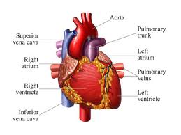
If the causative agent is Streptococcus viridans, the symptoms are generally mild with temperatures as prolonged loss of weight which may persist for several months. Contrary to this, the start can be very hectic with intermittent high fever, chills, fatigue, myalgia, arthralgia, headache, nausea, and vomiting.Objectively present the clinical findings of changes in the heart, changed the character's heartbeat, ekstratonova phenomenon, changing the character of noise or its appearance when it was not there before, and signs of congestive heart failure. Usually dominated by signs of mitral and aortic regurgitation. Splenomegaly was evident, and sometimes petechiae were present.

Neurological complications: embolism, cerebral abscesses, mikotične aneurysm and hemorrhage, manifested by signs of increased intracranial pressure, impaired senzorija tokalnim and neurological signs. Can be expected signs of systemic and pulmonary embolism.

Skin manifestations develop later in the disease, and are rarely seen in patients treated properly. These events are:

• Osler nodes (soft intradermal nodules on the side of your thumb palmarnoj)
• Janeway lesions (small erythematous or hemorrhagic painful lesions on the palms or soles)
• Splinter hemorrhages (linear lesions root of the nail)
• These lesions are probably caused by vasculitis, circulating antigen-antibody complexes.
Diagnosis:

The most important information to get the proper treatment haemoculture. Other laboratory data are of secondary importance are:

• Leukocytosis
• Hemolytic anemia
• rapidly
• microhematuria (a manifestation of immune complexes induced glomerulonephritis).
Blood culture done as soon as possible and in 3-5 times. In the first two blood cultures revealed the etiologic agent in 90% of cases. Previous antibacterial treatment to reduce blood culture positivity of 50-60%. Time sampling is not important since the expected constant bacteremia.

Echocardiography can reveal vegetation, identify their size, shape, location, and mobility as well as to determine the presence of valvular dysfunction. You can help in predicting complications emboličkih as lesions greater than 1 cm have a higher risk of embolization.

Prognosis and complications:

Forecast septic endocarditis is still serious. The mortality rate is 20-25%, and complications can be expected in 50-60% of children with documented disease.
The most common complications are:

• Congestive heart failure
• myocardial abscesses
• toxic myocarditis
• systemic embolization with signs of the CNS
• pulmonary embolism
• mikotične aneurysm
• ruptured sinus Valsalva
• acquired VSD
• disruptions in the implementation of pulse
Treatment:

Antibiotic treatment should be applied immediately after the diagnosis. A little delay could give progressive endocarditis with severe complications. One must keep a high level of bactericidal antibiotics long enough to eradicira microorganism that settled on the relatively avascular vegetation.Recommended duration of treatment within 4 to 6 weeks. The choice of antibiotics on the etiologic agent.

If it comes to Streptococcus viridans which is well susceptible to penicillin, then penicillin G is given in millions of doses, 200000-300000 IU / kg / day in 6 divided doses (every 4 hours), but not to exceed 20,000,000 for 24 hours . Alternatively given penicillin G plus gentaimicina iv 2-4 mg / kg / day in 3 divided doses, but not to exceed 80 mg / day.

If the cause of enterokokus which is less susceptible to penicillin, the preferred combination of iv Ampicillin 300 mg / kg / day in 4 divided doses with those that do not exceed 12 g / day, and gentamicin in the mentioned dose.

If the causative agent is Staphylococcus then semisintetski penicillins - oxacillin 200 mg / kg / day iv in 4 divided doses, but not to exceed 12 g / day.

Depending on the clinical response to therapy, in some cases it is necessary to prolong the treatment, and the highly sensitive infection with Streptococcus viridans may be recommended to shorten treatment time and include oral administration. If the heart is brought into a state of refractory congestive need the surgery, when the incorporation of artificial life-saving valve. Surgical replacement of the infected artificial valve has a large operational risk.

Prevention of septic endocarditis:

Surgical, dental procedures and the use of numerous instruments from different diagnostic and therapeutic procedures of mucosal damage caused by contamination of tissues with transient bacteremia that rarely persists for more than 15 minutes. These pathogens can settle on damaged, abnormal heart valves or heart defects in causing bacterial endocarditis or endarteritis.

Prophylactic use of antibiotics is recommended for patients who are at risk for developing endocarditis and underwent surgery that can cause bacteriemia. To avoid resistance to antibiotics used, prophylaxis is only used during the perioperative periods. 1 to 2 hours before surgery and 6 to 8 hours after.

State at risk for developing bacterial endocarditis:

• built-in artificial heart valves including bioprostetičke and homograft valves
• previous bacterial endocarditis, even in the absence of heart disease
• The majority of congenital cardiac malformations
• rheumatic valvular dysfunction or other etiology even after surgical intervention urađenog
• Hypertrophic Cardiomyopathy
• mitral valve prolapse with mitral regurgitation
Procedures that may cause bacteriemia:

• The majority of surgical procedures
• dental procedures that are known to cause gingival bleeding or mucous
Some conditions that are very close to the state where there is a risk of bacterial endocarditis but still bacterial endocarditis prophylaxis is not recommended:

• isolated ASD type sec.
• state after surgical correction without residue to 6 months after ASD correction type sec., VSD and patent ductus arteriosus
• mitral valve prolapse without mitral regurgitation - physiological, functional or inocentni noise
• previous Kawasaki-eva and rheumatic disease without valvular dysfunction
Some procedures that are close to those that can cause bacteriemia but still bacterial endocarditis prophylaxis is not recommended:

• stopping teeth chewing over the line
• Setting ortodontalnih appliances
• intraoral injections of anesthesia
• loss of primary teeth
• sectio caesarea
• uncomplicated vaginal delivery
The most numerous procedures that can cause bacteriemia smatotologije in the area. Poor oral hygiene, periodontal and periapical infections can cause bacteriemia even in the absence of dental procedures.People who have a risk of developing bacterial endocarditis should have the best possible oral health to eliminate a potential source of bakteriemije. Dentists should be reduced for patients with gingival inflammation stimulate brushing, rinsing with dezifencionim resources and professional teeth cleaning prior to performing routine dental procedures.

Before tooth extraction for 3 to 5 minutes to dry gum should be coated with CHLOREXIDINOM or povidone-iodine with because it has been proven that reduvira postekstrakcionu bacteriemia. If there is a dental procedure in the series then it is recommended to perform a seven-day intervals to reduce the potential risk of developing drug-resistant microorganisms, and patients without teeth may have the potential risk of ulceration due to bakteriemije mucosa that result from inadequately furnished prosthesis.

The most common cause of endocarditis following dental and surgical procedures in the upper respiratory tract (tonsils and / or adenoidectomy) is an α-hemolytic streptococcus and specific prophylaxis should be directed to the agent.
For a standard prophylactic regimen for all dental, oral surgical and surgical procedures in the upper respiratory tract is recommended amoxicillin. Amoxicillin is preferred over ampicillin and penicillin V due to its better absorption in the Gita, and the higher and more permanent level in the blood.

Granted to:

• AMOXICILLIN 50 mg / kg orally 1h before surgery, 25 mg / kg orally 6 to 8 hours after the initial dose. Being allergic to Amoxicillin / Penicillin or erythromycin given clyndamycin
• Erythromycin 20 mg / kg orally 2 h before surgery, 10 mg / kg orally 6 to 8 hours after the initial dose alternative
• CLYNDAMYCIN 10 mg / kg orally 1 hour before surgery, 5 mg / kg orally 6 to 8 hours after the initial dose. Total pediatric dose may exceed adult dose.
To determine the initial pediatric dose of amoxicillin can use the following scheme:

• below 15 kg body weight 750 mg amoxicillin
• 15 to 30 kg 1500 mg
• over 30 kg 3000 mg (adult dose)
• Doses below 1/2 of the initial dose.
Individuals who are unable to take medication orally recommended parenteral ampicillin.
Ampicillin 50 mg / kg im or iv 30 minutes before the procedure 25 mg / kg / m. or iv 6 hours after the initial dose. Being allergic to Ampicillin / Penicillin given Clyndamycin.
CLYNDAMYCIN 10 mg / kg iv 30 minutes prior to the procedure 5 mg / kg iv 6 hours after the initial dose.

Very high risk of developing endocarditis are those with artificial valves, surgically constructed systemic-pulmonary Santo, conduit and persons who have a history of earlier data prebolovanom endocarditis. In such persons develop endocarditis is associated with high mortality. In such persons recommended the combination of ampicillin and parenteral Gentamycin.

• Ampicillin 50 mg / kg im or iv 30 min. before surgery
• Gentamicin 2 mg / kg im or iv 30 min. before the procedure (not to exceed 80 mg). After 8 hours of repeating the initial dose regimen in full. Being allergic to Ampicillin / Penicillin given vancomycin.
• VANCOMYCIN 20 mg / kg iv perfusion for a period of 1 hour (to start 1 hour prior to surgery)
• Repeat dose is not necessary.
Bakteriemija can develop in surgical procedures and the use of different instruments for diagnostic and therapeutic purposes in genito-urinary and gastrointestinal tract. Bacterial endocarditis arising after such surgery usually is caused by Enterococcus foecalis. Antibiotic prophylaxis should be tailored to the agent. These requirements will meet the combination of:

• Ampicillin and Gentamycin as indicated above.
• If you are allergic to Ampicillin / Penicillin given combination VANCOMYCIN and Gentamycin.
• If you are low-risk patients in this group of patients is sufficient AMOXICILLIN orally.
• Tetracyclines and sulfonamides are not recommended in the prophylaxis of endocarditis.

Ventricular premature beats


Etiopathogenesis:

Ventricular ectopic strikes usually occur medikamenima carditis and intoxication. Bigeminy were found in digitalis intoxication.


The emergence of multi-focal ventricular prematurnih impact of different forms of a serious character.Characteristics of the ventricular premature stroke, in addition to a special form of time and lack of correlation with the activation of the atrium and full compensatory pause that follows them.

Series prematurnih ventricular stroke frequency of 120-180 with a minute if lucky, a reasonable doubt as to whether the present actual ventricular tachycardia or supraventricular tachycardia with aberrant intraventricular conduction. Dangers of ventricular tachycardia include:

• may result in congestive give or take longer if
• To go to the ventricular flutter or fibrillation and the result was instant death.
Treatment:

Xylocaine 1-2 mg / kg iv slowly with control of ECG and TA, Novocamid or 10 mg / kg / bw iv very slowly with control TA and ECG. If they are applied drug without effect, indicated electroconversion.

Right ventricular hypertrophy


The diagnosis of right ventricular hypertrophy has a far greater importance in children, than it is in adults. We found out that the cause of the changes in the electrical axis of the standard leads, and then determining hypertrophy can be followed by clinical context, determine the type and location in the heart of pataološkog state.


When right ventricular hypertrophy have the right axle standard deviation leads (S1 R3) and unipolar leads ekstremitetni show R or R1 in the AVR high-more than 6 mm, unipolar chest leads.

• Certain indications DVH exist when precordial leads V1 RS-deflection of the V6 (which points to the predominance of the action potential of the right ventricle through the entire prekordija.
• Type RS R1 deflection in V1 and V2, indicating a probable right ventricular hypertrophy, if R is greater than 15 mm, especially if R is greater than 10 mm.
• High initial RV1-2, with or without Q waves with small R and undoubtedly indicates DVH.
• Intrisikoidna R wave deflection of 0.04 seconds. or more in leads V1 and V2, reliable data for the DVH.
• Intervertirani T waves in leads desnogrudnim accompanied by ST-segment depression in conjunction with other changes in the chest drains indicates serious DVH.
• Indirect data for the DVH is taken high and pointed P2 indicates that the right atrial hypertrophy