Myocarditis
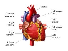
The heart consists of three layers: the endocardium (inner), myocardium (middle, muscular) and epicardium (outer).
Myocarditis is inflammation of the myocardium (middle layer of the heart wall). When this happens, the pump function of the heart becomes weak, because of swelling and damage to muscle cells. This damage can be caused by viruses, toxins, or occurs as an autoimmune process (immune cells to destroy your own heart cells).During viral infection, destruction and adverse remodeling of the heart can lead to permanent dysfunction of Commerce and the so-called. dilated cardiomyopathy.
Clinical picture can be different from asympthomatic to severe heart failure requiring heart transplantation, so that if myocarditis in the asymptomatic form, is equivocal for the diagnosis and timely treatment. Disease has many causes, and sometimes it happens that in case of sudden death does not reveal the culprit.
4-14 days after viral infection results in the formation of cells of the immune response of the organism. These cells attacking viruses, destroying myocardial cells (myocytes) in which the viruses are. Inadequate immune response may lead to a further multiplication of the virus and chronic destruction of heart cells, leading to dilated cardiomyopathy and heart failure.
Discovered the presence of so-called. antimiozinska autoantibodies, long after viral infection, which continues to destroy the myocytes and their presence indicates a poorer clinical prognosis. Over time, areas of necrosis of creating connective tissue. If a large myocardial involvement, there is a remodeling of cardiac cavities and their increase, leading to heart failure.
Focal inflammation which affects the conduction system can cause fatal heart arrhythmias that cause sudden death.
Sometimes myocarditis can give a picture of a heart attack.
Often occurs with myocarditis and pericarditis.
WHAT ARE THE CAUSES Myocarditis?
Infectious agents:
· Viruses: enteroviruses (Coxackie B) in> 50% of cases, adenoviruses, parvovirus B19, human herpes virus 6, influenza virus, echovirus, herpes simplex virus, varicella-zoster virus, hepatitis, Epstein-Barr virus and cytomegalovirus
· Bekterije: bekterije diphtheria, streptococcus, staphylococcus, brucella, salmonella, bacteria, Lyme disease carried by ticks
· Parasites: trichina, toxoplasmosis
Toxic causes:
· Some drugs can cause a hypersensitive reaction: clozapine, penicillin, ampicillin, hydrochlorothiazide, methyldopa, and sulfonamides
· Other drugs can destroy the heart cells: lithium, doxorubicin, cocaine, catecholamines, acetaminophen
· Toxins: lead, arsenic, carbon monoxide
· Toxins of some spiders, scorpions, wasps
· Air Therapy
Immune diseases:
· Systemic lupus, reumatoid arthritis, dermatomyositis, scleroderma, sarcoidosis, giant cell arteritis.
WHAT ARE THE SYMPTOMS Myocarditis?
Most patients have nonspecific symptoms such as fatigue, shortness of breath easily, and muscle aches. If challenged Coxackie B virus usually occur first stomach problems (diarrhea and abdominal cramps). When adenoviruses can occur sore throat, aching muscles and joints.
· A small number of patients have rapid and malignant course of disease with symptoms of heart failure.
· In about 20% of cases present fever.
· Heart pounding and skipping are present due to cardiac arrhythmias. Syncope (fainting) can occur if there are more difficult forms of arrhythmia or atrioventricular block. They are dangerous because they are a sign of poor prognosis, ie. risk of sudden cardiac death.
· One third of patients have chest pain. They describe them as a sharp stabbing chest, because it is often present, and pericarditis (inflammation of the pericardial sac). Pain may also occur below the breastbone in the projection of the stomach, which can mimic the pain of a heart attack.
· Difficulty breathing in effort is often present.
· Choking in the supine position is present if there is heart failure. The patient may fall and cardiogenic shock.
· In some patients the disease becomes chronic, and manifest only later with the expansion of cardiac cavities and the development of dilated cardiomyopathy.
HOW Myocarditis is diagnosed?
· Laboratory tests: troponin (elevated in one third of cases), leukocytes (raised in a quarter of cases), sedimentation (may be high), tients antibodies are sometimes found in immune-mediated myocarditis.
· X-ray of the heart and lungs: usually a normal cardiac silhouette, except when there is pericarditis or delay in the lungs due to heart failure. Sometimes there is a pericardial effusion in a handkerchief.
· Echocardiography: reduction of systolic and diastolic left ventricular function, regional or diffuse reduction in wall motion of the heart, possibly the presence of fluid in the pericardial cavity and sometimes the presence of thrombus in the left ventricle.
· MRI: a visible inflammation of areas of tissue.
· ECG: can be found sinus tachycardia, changes in ST segment, QRS complex, arrhythmias (SVES, VES), heart blocks.
· Isolation of virus from the blood.
· Sometimes it is necessary to do cardiac catheterization and endomyocardial biopsy (a catheter is accessed through the veins to the heart and takes a sample of heart tissue that is sent to the histopathological laboratory).
Pericarditis
Pericarditis is an inflammatory process that involves swelling and irritation of the pericardium, heart tissues.Pericardium has two layers, unutašnji which is attached to the heart muscle that surrounds the outer roots of the major blood vessels of the heart and through the ligament (connection) attaches the heart to surrounding organs: spinal column, diaphragm and other neighboring parts of the body. Between these two layers of pericardium is about 50 ml of fluid, an ultrafiltrate of blood plasma, and facilitates the movement of the heart.This liquid is drained through the lymphatic vessel in his right lung tissue.
Pericardium has three functions. The first is a mechanical function, prevents sudden dilation (expansion) of the heart, maintain a normal extension of commerce, forming an enclosed space with subatmosferskim atrial pressure, which helps fill with blood. Second, it protects the heart from external friction and prevent the spread of infection and malignancy of the heart. Thirdly, through its relationship with surrounding organs, pericardium fixed heart.
What happens in the pericardium?
During inflammation of the pericardium leads to accumulation of leukocytes. Often occurs and accumulation of fluid between two layers of the pericardium, the so-called. perikardnoj bag, which puts pressure on the heart and hinders its function. It can lead to creation of adhesions between the pericardium and the adjacent organs.
Pericarditis can be acute (lasting about 3 weeks) or chronic (lasting 6 months or longer).
WHAT CAUSES Pericarditis?
viruses, especially coxackie B and echovirus, adenoviruses, herpes viruses, citomegalivirus, influenza virus
bacteria: streptococci, staphylococci, pneumococci, Proteus, Escherichia coli, Pseudomonas
causative agent of Lyme disease (transmitted by ticks)
tuberculosis
inflammatory disorders, the so-called systemic diseases (arthritis-reumatoid common pericardial effusion, systemic lupus erythematosus, scleroderma (also with pleural effusion or constrictive pericarditis), rheumatic fever, sarcoidosis, etc.).
metabolic disorders (renal failure, reduced thyroid function, cholesterol pericarditis due to the high-cholesterol), low albumin levels.
after a heart attack affecting the entire wall of the heart, pericarditis usually occurs the second or third day. Is due to irritation of the pericardium partially damaged heart muscle.
aortic dissection
thoracic injuries
surgical procedure
Tumors
drugs (penicillin, doxorubicin, cyclophosphymid, procainamide, methyldopa, hydralazine, reserpine. Pericarditis caused by hypersensitivity reactions to the drug.
Valika radiotherapy doses of over 4000.
RECURRENT (RETURN) Pericarditis
It is the repetition of acute pericarditis. It can occur with or without accumulation of fluid (effusion) in perikardnoj bag and sometimes is associated with accumulation of fluid in the pulmonary tissue or changes in lung tissue. The reason for repeating pericarditis is not entirely clear, but it is assumed that it generates an autoimmune process. Responds well to nonsteroidal anti-inflammatory drugs and kortikosteride.
What are the symptoms Pericarditis?
Chest pain is the main symptom.
It can be: sharp, dull, or burning in the form of pressure in the chest.
intensity varies from very mild to severe
Pain occurs in the chest, could be disseminated in the left shoulder and back, increases when lying down, coughing and breathing air, a decrease in the sitting and leaning the body forward.
shortness of breath is often associated with pericardial tamponade (rapid accumulation of fluid in the sac perikardnoj)
fever may be present
weakness, fatigue
Island leg or stomach pain
HOW Pericarditis is diagnosed?
LABORATORY FINDINGS: acute pericarditis often elevated leukocyte count, C-reactive pritein, check the level of creatinine to assess renal function, do the analysis of cardiac enzymes to differentiate myocardial infarction, viral batteries; proteins in the blood, then testing in suspected tuberculosis; antistreptolizinski titer, rheumatoid factor, antinuclear antibodies, assessment of thyroid function.
ECG
X-ray of the heart and lungs: it shows an enlarged cardiac silhouette, if you have accumulated a lot of fluid in the pericardium. If there is a small amount of effusion, X-ray was normal.
ECHOCARDIOGRAPHY: shows the movement of cardiac walls, the size of cardiac cavities and the amount of fluid in the pericardial cavity.
CT
MRI
Pericarditis How is it treated?
inaction
used drugs that reduce inflammation and swelling and pain: nonsteroidal antiinflammatory drugs (Brufen), and if you need some stronger medication for pain.
Corticosteroids can be given if the disease does not respond to anti-rheumatic drugs, or if repeated often.
if it is a bacterial infection treated with antibiotics, and sometimes we need to give:
pericardiocentesis: puncture of the pericardium and fluid evacuation. There is a pericardial catheter drainage bag is gradual fluid from it.
Sometimes the therapy is resection of the pericardium, cut part of the pericardium
pericardiectomy is a surgical procedure in which the outer layer resecira pericardium. Performed with pericarditis konstriktivnog disruptive normal heart work.
What are the complications Pericarditis?
Constrictive pericarditis - inflammation of the pericardium after long and repeated chronic disease, pericardial thickening occurs, it creates scar tissue that SKVRCE, it is often the turbidity settle down calcium and to put pressure on the heart. The heart is the "shield" that prevents the normal spreading and taking blood from the venous system. Because we have a weakness of the right ventricle, there are islands on the feet, liver and spleen enlargement, fluid in the abdominal cavity. Later there is a weakness of the liver, blood in the veins stop the door, poured in the pulmonary maramici.Vremenom occurs and the weakness of the left heart, stagnation of blood in the lungs, decreased blood flow to internal organs, coldness in hands and feet, loss of consciousness due to decreased blood to the brain, fatigue and fatigue.
CAUSES: In addition to chronic pericarditis, causes include: tuberculosis, radiation therapy, uremia (due to renal failure), tumors after surgical interventions on the heart, and may be of unknown cause called.idiopathic.
TREATMENT: pericardiectomy, or surgical removal of the pericardium.
Pericardial tamponade - occurs when fluid accumulates rapidly in perikardnoj bag and then put pressure on the heart, thus preventing it from filling with blood. This means that less blood is pumped from the heart. The amount of pericardial fluid that is needed to reduce the filling pressure depends on the speed of accumulation of fluid and the elasticity of the pericardium. If fluid accumulates rapidly, a small amount of liquid such as 150 ml can cause a sudden pressure port in the pericardial cavity and significantly reduce the amount of blood that is pumped from the heart, causing a dramatic drop in blood pressure. If left untreated, ends fatally. If fluid accumulates slowly, 1000 ml of fluid will not significantly affect the amount of blood that takes chamber during diastole (expansion). This is due to the gradual spread of the pericardium.
SYMPTOMS: labored and rapid breathing, rapid heartbeat, and some people can appear cold extremities due to decreased blood flow. Other symptoms depend on the reasons that caused the tamponade.
CAUSES:
· Systemic malignant disease (weakness, fatigue, weight loss)
· Pericarditis-viral, bacterial, tuberculous, fungal (chest pain)
· Heart attack (chest pain)
· Anticoagulant therapy
· Diseases of connective tissue (muscle, bones and joints)
· Renal failure
· Recent heart surgery or intervention in coronary vessels
· Chest injury
· Recent implantation of a pacemaker or central venous catheter
· Aortic dissection (separation of the walls of the aorta)
· HIV
· Thoracic radiation therapy for malignant disease of the lungs or esophagus
· Tuberculosis
TREATMENT: cardiac tamponade is a medical emergency in medicine. The patient is hospitalized and monitored in intensive care.
The patient receives oxygen
infusions to compensate for the volume of fluid in blood vessels
drugs that enhance the strength of cardiac contraction (spasm)
it is urgent pericardiocentesis to drain fluid in the pericardium
in patients who have recurrence perkiarditisa Makes a pericardial-pleural shunt, ie the link between cardiac and pulmonary tissues to fluid in the pericardial sac drainaging lung tissue.
sometimes it is resection of the pericardium, ie removal of bags to prevent cardiac tamponade pericardium.
Endocarditis
Endocarditis is an infection of the inner layer of the heart called. endocardium.
Endocarditis occurs when bacteria or other germs spreading through the bloodstream from one place in the body, such as. mouth, heart attack damaged parts. If left untreated, can damage or destroy heart valves and lead to life-threatening complications.
Endocarditis is rare in people with healthy hearts. In people who have damaged heart valves, artificial valves or other cardiac defects, there is an increased risk of developing endocarditis.
What are the symptoms Endocarditis?
Endocarditis may develop gradually or abruptly, depending on what is causing the infection and whether there is a heart defect.
The symptoms are:
fever
chills
fatigue
muscle and joint pain
night znojenjene
lack of air
cough
Islands of legs and abdomen
sudden weight loss
pain under left rib cage
Osler nodes-tiny red dots under the skin sore fingertips
petechiae-point bleeding under the skin, mucous membranes in the mouth and whites of the eye
embolism (parts of damaged tissue to detach and move through the bloodstream, and clog an artery of the) brain and neurological deficit occurs, the eye - vision loss, signs of systemic septic embolism (eg, pulmonary infection if endokaridits right in the heart)
Sometimes the appearance of blood in the urine.
Of these characters may appear only a few, perhaps only two.
WHAT ARE THE CAUSES Endocarditis?
Bacteria are the most common causes of endocarditis, but it may be a fungus and other microorganisms.
Sometimes the culprit is one of the many bacteria that live in your mouth, throat or other parts of the body. In other cases, bacteria can enter the body during tooth brushing, chewing food, especially if the teeth and gums are in poor condition. The causative agent can be spread from sites of infection, eg. wounds on the skin, inflammatory bowel disease, from entering the body through sexual contact etc.. It also increased the possibility of bacteria entering the body through a needle in drug users, tattooing, body-piersing a following by people who have a catheter in the bladder, in patients with implanted pacemaker. Some dental procedures can damage the gums allow bacteria to enter the bloodstream.
Your immune system destroys the bacteria that enter the bloodstream. Even if they reach the heart, must not cause an infection.
Most people who develop endocarditis have a damaged heart valves. Endocardium is rugged making it an ideal place for the establishment and growth of bacteria.
WHO HAS increased risk of Endocarditis?
People with artificial heart valves. The risk of endocarditis is increased in the first year after valve implantation.
People with congenital heart disease.
Individuals who have had endocarditis. Previous endocarditis damaged heart tissue or valves, and a suitable place for the redevelopment of endocarditis.
People with damaged heart valves, rheumatic fever or infection can damage heart valves to make them suitable terrain for the development of endocarditis.
Injecting drug users
People with heart transplant who have problems with heart valves.
How is it diagnosed
Work by biochemical analysis of blood
is the identification of pathogens from the blood in order to include the appropriate antibiotic to which the germ-sensitive
ECG
echocardiography
X-ray of the heart and lungs
heart catheterization
CT or MRI of the brain, lungs, abdomen and others. if there is suspected spread of infection to other organs.
What is the treatment Endocarditis?
· These patients are hospitalized and begins to empirical antibiotic therapy.
· Gives the oxygen
· Provide the drugs for heart failure
· When the situation improves, assesses the need for surgical treatment.
Endocarditis Prophylaxis
Is required for all persons with an increased risk before one of the following medical interventions:
dental procedures
medical interventions for which there is damage to the lining of the airways
procedures on infected skin and musculo-skeletal tissue
prophylaxis is not required for gynecological and urological procedures and interventions in the digestive tract.
Since antibiotics before dental procedures or interventions on airway mucosa taken:
Oral Amoxicillin 2 g, 30-60 min before procedure
Allergy to penicillin is taken: 2g Cephalexin or Clindamycin 600mg Azithromycin or Clarithromycin 500 mg or 500 mg 30-60 minutes before procedure
If people can not take oral medication is given: Ceftriaxon 1g of muscle or vein
Allergy to penicillin is given: Ceftriaxon 1g into a muscle or vein or Clindamycin 600 mg in a muscle or vein.
What are the complications Endocarditis?
Infarcts of other organs and tissues: heart, brain, spleen, intestine, kidney, extremities. They are formed by joining the masses of bacteria and cell fragments of the affected valve (the vegetation) and reach the bloodstream to other organs and tissues and deplete them
Infections of other parts of the body: abscesses caused the brain, heart, lung, kidney, spleen, liver, heart (arrhythmia occurs)
Pericarditis
Heart failure due to valvular damage functions
Kidney failure
If left untreated leads to death.
Prognosis
Increased mortality occurs in older individuals, then if the infection has affected the aortic valve, if there was a development of heart failure, complications of the brain, and if a person suffers from other serious diseases such as diabetes.
Mortality depends on the cause of endocarditis and appears to be greatest in Staphylococcus aureus.
Mortality was higher in patients with prosthetic heart valves. More than 50% of infection occurs within 2 months after valve surgery.
Useful
information about health and healthy diet you can find on: